Understanding Soft Tissue Evaluations in Diagnosis

Understanding Soft Tissue Evaluations in Diagnosis

Brief overview of orthodontic treatment for kids and the importance of imaging methods in diagnosis and treatment planning

Sure! Here's a short essay on the topic "Understanding Soft Tissue Evaluations in Diagnosis: Overview of the Role of Soft Tissues in Facial Aesthetics and Function, Highlighting Their Impact on Orthodontic Outcomes."


Orthodontic treatment can help improve your child's smile Dental braces for children United States.

In the realm of orthodontics, the evaluation of soft tissues plays a crucial role in both diagnosis and treatment planning. Soft tissues, which include the skin, muscles, and other connective tissues of the face, significantly influence facial aesthetics and function. Understanding their impact is essential for achieving optimal orthodontic outcomes.


Firstly, soft tissues contribute to the overall harmony and balance of facial features. The position and contour of the lips, cheeks, and even the nose are influenced by the underlying skeletal structure as well as the soft tissues themselves. For instance, a well-aligned jaw not only ensures proper dental function but also enhances the aesthetic appeal of the smile and profile. Orthodontists must consider how changes in tooth and jaw alignment will affect these soft tissue structures to ensure that the final result is both functional and aesthetically pleasing.


Moreover, soft tissues play a dynamic role in facial expressions and communication. The muscles of facial expression, which are embedded within the soft tissues, allow us to smile, frown, and convey a wide range of emotions. Any alteration in the underlying skeletal structure due to orthodontic treatment can affect these muscle functions. Therefore, a comprehensive soft tissue evaluation helps orthodontists anticipate and mitigate potential issues, ensuring that the patient's ability to express themselves remains unhindered.


In terms of function, soft tissues are integral to mastication, speech, and even breathing. The position of the tongue, for example, is influenced by the surrounding soft tissues and can impact speech clarity and swallowing efficiency. Orthodontists must evaluate these relationships to prevent functional impairments post-treatment.


Additionally, the interaction between soft tissues and hard tissues is a critical factor in the stability of orthodontic outcomes. Retainers and other post-treatment appliances are often necessary to maintain the new alignment of teeth, but the pressure exerted by surrounding soft tissues can influence how well these results are sustained over time.


In conclusion, a thorough understanding of soft tissue evaluations in diagnosis is indispensable in orthodontics. It not only enhances the aesthetic outcomes but also ensures functional integrity and long-term stability. By meticulously assessing and considering the role of soft tissues, orthodontists can craft treatment plans that are both effective and holistic, ultimately leading to satisfied patients with beautiful, healthy smiles.

Understanding soft tissue evaluations in diagnosis is crucial for various medical and dental fields, as it helps practitioners gain a comprehensive view of a patient's condition. Detailed examination techniques for assessing soft tissue profiles play a significant role in this process. These techniques encompass visual inspection, photographic analysis, and cephalometric radiographs, each contributing unique insights into the patient's soft tissue structure and function.


Visual inspection is the most straightforward and initial method used in soft tissue evaluation. It involves a thorough, unaided examination of the patient's external features, looking for any abnormalities, asymmetries, or irregularities in the soft tissues. This method relies heavily on the clinician's experience and observational skills. It allows for the immediate identification of obvious issues such as swelling, discoloration, or deformities. However, visual inspection alone may not provide a complete picture, as it is limited to surface-level observations.


Photographic analysis introduces a more detailed and objective approach to soft tissue evaluation. By capturing images of the patient from various angles, clinicians can analyze the soft tissue profile more precisely. Digital photography allows for magnification, comparison over time, and detailed documentation of the patient's condition. This technique is particularly useful in tracking changes in soft tissue structure following treatment or in planning surgical interventions. Moreover, photographic analysis can be enhanced with specialized software that measures dimensions and angles, providing quantitative data to support qualitative observations.


Cephalometric radiographs represent the most sophisticated technique in soft tissue evaluation. These X-ray images provide a detailed view of the underlying bone structure as well as the overlying soft tissues. By analyzing cephalometric radiographs, clinicians can assess the relationship between the hard and soft tissues, identify any discrepancies in growth patterns, and plan orthognathic surgeries with greater precision. This technique allows for a three-dimensional understanding of the patient's anatomy, facilitating more accurate diagnoses and treatment plans.


In conclusion, the detailed examination techniques for assessing soft tissue profiles-visual inspection, photographic analysis, and cephalometric radiographs-are indispensable tools in the diagnosis and treatment planning process. Each method offers unique advantages, and when used in combination, they provide a comprehensive evaluation of the patient's soft tissue condition. This multifaceted approach ensures that clinicians can make well-informed decisions, leading to more effective and personalized patient care.

Description of the benefits and limitations of each imaging method, including factors such as radiation exposure, image quality, and cost

Certainly! Understanding soft tissue evaluations in pediatric orthodontics is crucial for achieving successful treatment outcomes. Soft tissues, including the lips, cheeks, and tongue, significantly influence the development and alignment of teeth and jaws. In pediatric patients, common soft tissue discrepancies can greatly impact orthodontic diagnosis and treatment planning.


One prevalent discrepancy is the incompetent lip seal, where a child cannot close their lips together at rest. This condition is often associated with mouth breathing, which can lead to a narrow maxillary arch, proclined upper incisors, and an open bite tendency. In orthodontic treatment, addressing the underlying cause of mouth breathing, such as nasal obstruction, is essential. Additionally, orthodontic appliances may be used to expand the maxillary arch and correct the inclination of the incisors.


Another common issue is abnormal tongue posture or function. A low tongue posture or a tongue thrust habit can exert pressure on the anterior teeth, leading to spacing or proclination. During orthodontic evaluation, observing tongue habits and their impact on dental alignment is vital. Treatment may involve myofunctional therapy to retrain the tongue's resting position and swallowing pattern, alongside orthodontic appliances to correct tooth positioning.


Cheek and lip pressure also play a significant role in dental development. Excessive pressure from these soft tissues can contribute to dental crowding or misalignment. Evaluating the balance between these forces is essential in orthodontic diagnosis. Treatment strategies may include the use of appliances that redistribute pressure or techniques that modify soft tissue behavior to create a more favorable environment for tooth alignment.


In conclusion, a thorough evaluation of soft tissue discrepancies in pediatric patients is fundamental in orthodontic diagnosis. Understanding these factors allows orthodontists to develop comprehensive treatment plans that not only address dental misalignments but also consider the dynamic interplay between soft tissues and dental structures. This holistic approach ensures more stable and aesthetically pleasing outcomes in pediatric orthodontic treatment.

Description of the benefits and limitations of each imaging method, including factors such as radiation exposure, image quality, and cost

Discussion of the role of digital imaging technologies in modern orthodontics, including the use of 3D imaging and computer-aided design and manufacturing (CAD/CAM) systems

When it comes to dental and orthodontic treatment, soft tissue evaluations play a crucial role in creating effective and comprehensive treatment plans. Soft tissues, which include the lips, cheeks, tongue, and other facial muscles, significantly influence both the function and aesthetics of the dentofacial complex. Understanding how to integrate these evaluations into treatment plans, especially considering growth and development, can lead to better outcomes and patient satisfaction.


Firstly, it's essential to recognize that soft tissue evaluations go beyond mere observation. They require a detailed analysis of the patient's profile, facial symmetry, lip position, and the relationship between the lips and teeth. This analysis helps practitioners predict how treatment will affect facial aesthetics and function. By incorporating these evaluations early in the diagnostic process, clinicians can develop a more holistic view of the patient's needs.


One effective strategy is to use photographic and radiographic assessments in conjunction with clinical examinations. Photographs provide a visual record of the patient's facial features and can be invaluable for tracking changes over time. Lateral cephalometric radiographs offer a detailed look at the skeletal and soft tissue structures, allowing for a more accurate diagnosis and treatment planning.


Considering growth and development is particularly important in pediatric and adolescent patients. The dynamic nature of growth means that treatment plans must be flexible and adaptable. For younger patients, growth modification techniques can be employed to guide jaw development and improve soft tissue relationships. This proactive approach can prevent more complex issues from arising in the future.


In adult patients, where growth is no longer a factor, soft tissue considerations still play a vital role. Treatment plans might focus more on achieving harmony between the teeth, jaws, and facial features. For instance, in cases where extractions are necessary, careful planning is required to ensure that the resulting changes in tooth position do not adversely affect the patient's profile or lip support.


Another strategy is to involve patients in the decision-making process. Educating patients about the importance of soft tissue evaluations and how they influence treatment outcomes can lead to greater cooperation and satisfaction. When patients understand the rationale behind certain treatment decisions, they are more likely to adhere to the recommended plan.


Lastly, continuous education and training for practitioners are crucial. The field of orthodontics and dentistry is constantly evolving, with new techniques and technologies emerging regularly. Staying abreast of the latest research and advancements in soft tissue evaluations ensures that practitioners can offer the best possible care to their patients.


In conclusion, integrating soft tissue evaluations into comprehensive treatment plans is a multifaceted approach that requires careful consideration of growth and development, the use of advanced diagnostic tools, patient education, and ongoing professional development. By adopting these strategies, practitioners can enhance the aesthetic and functional outcomes of their treatments, leading to happier and more satisfied patients.

Overview of the importance of proper image interpretation and analysis in orthodontic treatment planning, including the use of landmarks, measurements, and tracings

In the realm of orthodontics, the assessment of soft tissue plays a pivotal role in diagnosing and treating various issues, particularly in children. This essay delves into the significance of soft tissue evaluations, highlighting their application through case studies.


Firstly, it's essential to comprehend the role of soft tissue in orthodontic diagnosis. Soft tissues, including the lips, cheeks, and tongue, exert forces on the teeth and jaw, influencing their positioning. Evaluating these tissues allows orthodontists to identify potential causes of malocclusions, such as improper tongue posture or lip tension.


One case study involves a 10-year-old boy presenting with a Class II malocclusion characterized by an overjet. Through soft tissue evaluation, the orthodontist identified excessive lip tension as a contributing factor. By incorporating myofunctional therapy to address the lip posture, alongside traditional orthodontic treatment, the boy achieved significant improvement in his bite alignment.


Another case study features an 8-year-old girl with a Class III malocclusion, exhibiting an anterior crossbite. Soft tissue assessment revealed a forward tongue posture, exerting pressure on the lower incisors. Utilizing habit-breaking appliances and tongue retraining exercises, the orthodontist successfully corrected the crossbite and improved the girl's facial profile.


Furthermore, soft tissue evaluation aids in diagnosing functional issues such as mouth breathing or tongue thrust, which can exacerbate orthodontic problems. In a case involving a 9-year-old boy with a narrow maxillary arch and spacing between the upper incisors, soft tissue assessment revealed habitual mouth breathing due to nasal obstruction. Collaborating with an ENT specialist to address the nasal issue and incorporating orthodontic treatment to expand the maxillary arch yielded favorable outcomes.


In conclusion, soft tissue evaluations are indispensable in orthodontic diagnosis and treatment planning, especially in children. Through case studies, we witness the profound impact of assessing soft tissues on achieving successful orthodontic outcomes. By integrating soft tissue evaluations into clinical practice, orthodontists can address underlying functional issues and optimize treatment outcomes for young patients.

Explanation of the role of orthodontic imaging in monitoring treatment progress and evaluating treatment outcomes

In the realm of pediatric orthodontics, understanding soft tissue evaluations is crucial for accurate diagnosis and effective treatment planning. Recent research and advancements in soft tissue analysis techniques have significantly enhanced our ability to assess and interpret these evaluations.


Traditionally, orthodontic assessments have relied heavily on hard tissue analysis, such as cephalometric radiographs, to evaluate skeletal and dental structures. However, the role of soft tissues-including the lips, cheeks, and tongue-in facial aesthetics and function cannot be overstated. Soft tissue evaluations provide insights into the dynamic interplay between facial muscles and underlying skeletal structures, which is particularly important in growing children.


One of the notable advancements in this field is the integration of three-dimensional (3D) imaging technologies. Techniques such as 3D photogrammetry and cone-beam computed tomography (CBCT) allow for a more comprehensive and detailed analysis of soft tissues. These methods offer high-resolution images that capture the nuances of facial morphology, enabling orthodontists to make more informed decisions.


Moreover, software advancements have facilitated the development of automated soft tissue analysis tools. These tools can quickly process 3D images to generate detailed reports, highlighting areas of concern and predicting treatment outcomes. Such automation not only saves time but also enhances the accuracy of diagnoses.


Another significant area of research is the use of machine learning algorithms to analyze soft tissue patterns. By training algorithms on large datasets of images, researchers can identify subtle correlations between soft tissue characteristics and orthodontic issues. This approach allows for personalized treatment plans tailored to the unique facial features of each patient.


Additionally, the integration of soft tissue analysis into digital smile design (DSD) has revolutionized the way orthodontists approach aesthetic treatments. DSD involves creating a virtual mock-up of the patient's smile, taking into account both hard and soft tissue elements. This technique helps in visualizing the potential outcomes of orthodontic interventions, thereby improving patient communication and satisfaction.


In conclusion, the review of current research and advancements in soft tissue analysis techniques underscores their growing importance in pediatric orthodontics. By embracing these innovations, orthodontists can achieve more precise diagnoses, develop tailored treatment plans, and ultimately enhance the aesthetic and functional outcomes for their young patients.

Human lower jaw viewed from the left

The jaws are a pair of opposable articulated structures at the entrance of the mouth, typically used for grasping and manipulating food. The term jaws is also broadly applied to the whole of the structures constituting the vault of the mouth and serving to open and close it and is part of the body plan of humans and most animals.

Arthropods

[edit]
The mandibles of a bull ant

In arthropods, the jaws are chitinous and oppose laterally, and may consist of mandibles or chelicerae. These jaws are often composed of numerous mouthparts. Their function is fundamentally for food acquisition, conveyance to the mouth, and/or initial processing (mastication or chewing). Many mouthparts and associate structures (such as pedipalps) are modified legs.

Vertebrates

[edit]

In most vertebrates, the jaws are bony or cartilaginous and oppose vertically, comprising an upper jaw and a lower jaw. The vertebrate jaw is derived from the most anterior two pharyngeal arches supporting the gills, and usually bears numerous teeth.

Jaws of a great white shark

Fish

[edit]
Moray eels have two sets of jaws: the oral jaws that capture prey and the pharyngeal jaws that advance into the mouth and move prey from the oral jaws to the esophagus for swallowing.

The vertebrate jaw probably originally evolved in the Silurian period and appeared in the Placoderm fish which further diversified in the Devonian. The two most anterior pharyngeal arches are thought to have become the jaw itself and the hyoid arch, respectively. The hyoid system suspends the jaw from the braincase of the skull, permitting great mobility of the jaws. While there is no fossil evidence directly to support this theory, it makes sense in light of the numbers of pharyngeal arches that are visible in extant jawed vertebrates (the Gnathostomes), which have seven arches, and primitive jawless vertebrates (the Agnatha), which have nine.

The original selective advantage offered by the jaw may not be related to feeding, but rather to increased respiration efficiency.[1] The jaws were used in the buccal pump (observable in modern fish and amphibians) that pumps water across the gills of fish or air into the lungs in the case of amphibians. Over evolutionary time the more familiar use of jaws (to humans), in feeding, was selected for and became a very important function in vertebrates. Many teleost fish have substantially modified jaws for suction feeding and jaw protrusion, resulting in highly complex jaws with dozens of bones involved.[2]

Amphibians, reptiles, and birds

[edit]

The jaw in tetrapods is substantially simplified compared to fish. Most of the upper jaw bones (premaxilla, maxilla, jugal, quadratojugal, and quadrate) have been fused to the braincase, while the lower jaw bones (dentary, splenial, angular, surangular, and articular) have been fused together into a unit called the mandible. The jaw articulates via a hinge joint between the quadrate and articular. The jaws of tetrapods exhibit varying degrees of mobility between jaw bones. Some species have jaw bones completely fused, while others may have joints allowing for mobility of the dentary, quadrate, or maxilla. The snake skull shows the greatest degree of cranial kinesis, which allows the snake to swallow large prey items.

Mammals

[edit]

In mammals, the jaws are made up of the mandible (lower jaw) and the maxilla (upper jaw). In the ape, there is a reinforcement to the lower jaw bone called the simian shelf. In the evolution of the mammalian jaw, two of the bones of the jaw structure (the articular bone of the lower jaw, and quadrate) were reduced in size and incorporated into the ear, while many others have been fused together.[3] As a result, mammals show little or no cranial kinesis, and the mandible is attached to the temporal bone by the temporomandibular joints. Temporomandibular joint dysfunction is a common disorder of these joints, characterized by pain, clicking and limitation of mandibular movement.[4] Especially in the therian mammal, the premaxilla that constituted the anterior tip of the upper jaw in reptiles has reduced in size; and most of the mesenchyme at the ancestral upper jaw tip has become a protruded mammalian nose.[5]

Sea urchins

[edit]

Sea urchins possess unique jaws which display five-part symmetry, termed the Aristotle's lantern. Each unit of the jaw holds a single, perpetually growing tooth composed of crystalline calcium carbonate.

See also

[edit]
  • Muscles of mastication
  • Otofacial syndrome
  • Predentary
  • Prognathism
  • Rostral bone

References

[edit]
  1. ^ Smith, M.M.; Coates, M.I. (2000). "10. Evolutionary origins of teeth and jaws: developmental models and phylogenetic patterns". In Teaford, Mark F.; Smith, Moya Meredith; Ferguson, Mark W.J. (eds.). Development, function and evolution of teeth. Cambridge: Cambridge University Press. p. 145. ISBN 978-0-521-57011-4.
  2. ^ Anderson, Philip S.L; Westneat, Mark (28 November 2006). "Feeding mechanics and bite force modelling of the skull of Dunkleosteus terrelli, an ancient apex predator". Biology Letters. pp. 77–80. doi:10.1098/rsbl.2006.0569. PMC 2373817. PMID 17443970. cite web: Missing or empty |url= (help)
  3. ^ Allin EF (December 1975). "Evolution of the mammalian middle ear". J. Morphol. 147 (4): 403–37. doi:10.1002/jmor.1051470404. PMID 1202224. S2CID 25886311.
  4. ^ Wright, Edward F. (2010). Manual of temporomandibular disorders (2nd ed.). Ames, Iowa: Wiley-Blackwell. ISBN 978-0-8138-1324-0.
  5. ^ Higashiyama, Hiroki; Koyabu, Daisuke; Hirasawa, Tatsuya; Werneburg, Ingmar; Kuratani, Shigeru; Kurihara, Hiroki (November 2, 2021). "Mammalian face as an evolutionary novelty". PNAS. 118 (44): e2111876118. Bibcode:2021PNAS..11811876H. doi:10.1073/pnas.2111876118. PMC 8673075. PMID 34716275.
[edit]
  • Media related to Jaw bones at Wikimedia Commons
  • Jaw at the U.S. National Library of Medicine Medical Subject Headings (MeSH)


Malocclusion
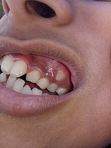
Malocclusion in 10-year-old girl
Specialty Dentistry Edit this on Wikidata

In orthodontics, a malocclusion is a misalignment or incorrect relation between the teeth of the upper and lower dental arches when they approach each other as the jaws close. The English-language term dates from 1864;[1] Edward Angle (1855–1930), the "father of modern orthodontics",[2][3][need quotation to verify] popularised it. The word derives from mal- 'incorrect' and occlusion 'the manner in which opposing teeth meet'.

The malocclusion classification is based on the relationship of the mesiobuccal cusp of the maxillary first molar and the buccal groove of the mandibular first molar.  If this molar relationship exists, then the teeth can align into normal occlusion. According to Angle, malocclusion is any deviation of the occlusion from the ideal.[4] However, assessment for malocclusion should also take into account aesthetics and the impact on functionality. If these aspects are acceptable to the patient despite meeting the formal definition of malocclusion, then treatment may not be necessary. It is estimated that nearly 30% of the population have malocclusions that are categorised as severe and definitely benefit from orthodontic treatment.[5]

Causes

[edit]

The aetiology of malocclusion is somewhat contentious, however, simply put it is multifactorial, with influences being both genetic[6][unreliable source?] and environmental.[7] Malocclusion is already present in one of the Skhul and Qafzeh hominin fossils and other prehistoric human skulls.[8][9] There are three generally accepted causative factors of malocclusion:

  • Skeletal factors – the size, shape and relative positions of the upper and lower jaws. Variations can be caused by environmental or behavioral factors such as muscles of mastication, nocturnal mouth breathing, and cleft lip and cleft palate.
  • Muscle factors – the form and function of the muscles that surround the teeth.  This could be impacted by habits such as finger sucking, nail biting, pacifier and tongue thrusting[10]
  • Dental factors – size of the teeth in relation to the jaw, early loss of teeth could result in spacing or mesial migration causing crowding, abnormal eruption path or timings, extra teeth (supernumeraries), or too few teeth (hypodontia)

There is not one single cause of malocclusion, and when planning orthodontic treatment it is often helpful to consider the above factors and the impact they have played on malocclusion. These can also be influenced by oral habits and pressure resulting in malocclusion.[11][12]

Behavioral and dental factors

[edit]

In the active skeletal growth,[13] mouthbreathing, finger sucking, thumb sucking, pacifier sucking, onychophagia (nail biting), dermatophagia, pen biting, pencil biting, abnormal posture, deglutition disorders and other habits greatly influence the development of the face and dental arches.[14][15][16][17][18] Pacifier sucking habits are also correlated with otitis media.[19][20] Dental caries, periapical inflammation and tooth loss in the deciduous teeth can alter the correct permanent teeth eruptions.

Primary vs. secondary dentition

[edit]

Malocclusion can occur in primary and secondary dentition.

In primary dentition malocclusion is caused by:

  • Underdevelopment of the dentoalvelor tissue.
  • Over development of bones around the mouth.
  • Cleft lip and palate.
  • Overcrowding of teeth.
  • Abnormal development and growth of teeth.

In secondary dentition malocclusion is caused by:

  • Periodontal disease.
  • Overeruption of teeth.[21]
  • Premature and congenital loss of missing teeth.

Signs and symptoms

[edit]

Malocclusion is a common finding,[22][23] although it is not usually serious enough to require treatment. Those who have more severe malocclusions, which present as a part of craniofacial anomalies, may require orthodontic and sometimes surgical treatment (orthognathic surgery) to correct the problem.

The ultimate goal of orthodontic treatment is to achieve a stable, functional and aesthetic alignment of teeth which serves to better the patient's dental and total health.[24] The symptoms which arise as a result of malocclusion derive from a deficiency in one or more of these categories.[25]

The symptoms are as follows:

  • Tooth decay (caries): misaligned teeth will make it more difficult to maintain oral hygiene. Children with poor oral hygiene and diet will be at an increased risk.
  • Periodontal disease: irregular teeth would hinder the ability to clean teeth meaning poor plaque control. Additionally, if teeth are crowded, some may be more buccally or lingually placed, there will be reduced bone and periodontal support. Furthermore, in Class III malocclusions, mandibular anterior teeth are pushed labially which contributes to gingival recession and weakens periodontal support.
  • Trauma to anterior teeth: Those with an increased overjet are at an increased risk of trauma. A systematic review found that an overjet of greater than 3mm will double the risk of trauma.
  • Masticatory function: people with anterior open bites, large increased & reverse overjet and hypodontia will find it more difficult to chew food.
  • Speech impairment: a lisp is when the incisors cannot make contact, orthodontics can treat this. However, other forms of misaligned teeth will have little impact on speech and orthodontic treatment has little effect on fixing any problems.  
  • Tooth impaction: these can cause resorption of adjacent teeth and other pathologies for example a dentigerous cyst formation.  
  • Psychosocial wellbeing: malocclusions of teeth with poor aesthetics can have a significant effect on self-esteem.

Malocclusions may be coupled with skeletal disharmony of the face, where the relations between the upper and lower jaws are not appropriate. Such skeletal disharmonies often distort sufferer's face shape, severely affect aesthetics of the face, and may be coupled with mastication or speech problems. Most skeletal malocclusions can only be treated by orthognathic surgery.[citation needed]

Classification

[edit]

Depending on the sagittal relations of teeth and jaws, malocclusions can be divided mainly into three types according to Angle's classification system published 1899. However, there are also other conditions, e.g. crowding of teeth, not directly fitting into this classification.

Many authors have tried to modify or replace Angle's classification. This has resulted in many subtypes and new systems (see section below: Review of Angle's system of classes).

A deep bite (also known as a Type II Malocclusion) is a condition in which the upper teeth overlap the lower teeth, which can result in hard and soft tissue trauma, in addition to an effect on appearance.[26] It has been found to occur in 15–20% of the US population.[27]

An open bite is a condition characterised by a complete lack of overlap and occlusion between the upper and lower incisors.[28] In children, open bite can be caused by prolonged thumb sucking.[29] Patients often present with impaired speech and mastication.[30]

Overbites

[edit]

This is a vertical measurement of the degree of overlap between the maxillary incisors and the mandibular incisors. There are three features that are analysed in the classification of an overbite:

  • Degree of overlap: edge to edge, reduced, average, increased
  • Complete or incomplete: whether there is contact between the lower teeth and the opposing teeth/tissue (hard palate or gingivae) or not.
  • Whether contact is traumatic or atraumatic

An average overbite is when the upper anterior teeth cover a third of the lower teeth. Covering less than this is described as ‘reduced’ and more than this is an ‘increased’ overbite. No overlap or contact is considered an ‘anterior open bite’.[25][31][32]

Angle's classification method

[edit]
Class I with severe crowding and labially erupted canines
Class II molar relationship

Edward Angle, who is considered the father of modern orthodontics, was the first to classify malocclusion. He based his classifications on the relative position of the maxillary first molar.[33] According to Angle, the mesiobuccal cusp of the upper first molar should align with the buccal groove of the mandibular first molar. The teeth should all fit on a line of occlusion which, in the upper arch, is a smooth curve through the central fossae of the posterior teeth and cingulum of the canines and incisors, and in the lower arch, is a smooth curve through the buccal cusps of the posterior teeth and incisal edges of the anterior teeth. Any variations from this resulted in malocclusion types. It is also possible to have different classes of malocclusion on left and right sides.

  • Class I (Neutrocclusion): Here the molar relationship of the occlusion is normal but the incorrect line of occlusion or as described for the maxillary first molar, but the other teeth have problems like spacing, crowding, over or under eruption, etc.
  • Class II (Distocclusion (retrognathism, overjet, overbite)): In this situation, the mesiobuccal cusp of the upper first molar is not aligned with the mesiobuccal groove of the lower first molar. Instead it is anterior to it. Usually the mesiobuccal cusp rests in between the first mandibular molars and second premolars. There are two subtypes:
    • Class II Division 1: The molar relationships are like that of Class II and the anterior teeth are protruded.
    • Class II Division 2: The molar relationships are Class II but the central are retroclined and the lateral teeth are seen overlapping the centrals.
  • Class III: (Mesiocclusion (prognathism, anterior crossbite, negative overjet, underbite)) In this case the upper molars are placed not in the mesiobuccal groove but posteriorly to it. The mesiobuccal cusp of the maxillary first molar lies posteriorly to the mesiobuccal groove of the mandibular first molar. Usually seen as when the lower front teeth are more prominent than the upper front teeth. In this case the patient very often has a large mandible or a short maxillary bone.

Review of Angle's system of classes and alternative systems

[edit]

A major disadvantage of Angle's system of classifying malocclusions is that it only considers two dimensions along a spatial axis in the sagittal plane in the terminal occlusion, but occlusion problems can be three-dimensional. It does not recognise deviations in other spatial axes, asymmetric deviations, functional faults and other therapy-related features.

Angle's classification system also lacks a theoretical basis; it is purely descriptive. Its much-discussed weaknesses include that it only considers static occlusion, it does not account for the development and causes (aetiology) of occlusion problems, and it disregards the proportions (or relationships in general) of teeth and face.[34] Thus, many attempts have been made to modify the Angle system or to replace it completely with a more efficient one,[35] but Angle's classification continues be popular mainly because of its simplicity and clarity.[citation needed]

Well-known modifications to Angle's classification date back to Martin Dewey (1915) and Benno Lischer (1912, 1933). Alternative systems have been suggested by, among others, Simon (1930, the first three-dimensional classification system), Jacob A. Salzmann (1950, with a classification system based on skeletal structures) and James L. Ackerman and William R. Proffit (1969).[36]

Incisor classification

[edit]

Besides the molar relationship, the British Standards Institute Classification also classifies malocclusion into incisor relationship and canine relationship.

  • Class I: The lower incisor edges occlude with or lie immediately below the cingulum plateau of the upper central incisors
  • Class II: The lower incisor edges lie posterior to the cingulum plateau of the upper incisors
    • Division 1 – the upper central incisors are proclined or of average inclination and there is an increase in overjet
    • Division 2 – The upper central incisors are retroclined. The overjet is usually minimal or may be increased.
  • Class III: The lower incisor edges lie anterior to the cingulum plateau of the upper incisors. The overjet is reduced or reversed.

Canine relationship by Ricketts

[edit]
  • Class I: Mesial slope of upper canine coincides with distal slope of lower canine
  • Class II: Mesial slope of upper canine is ahead of distal slope of lower canine
  • Class III: Mesial slope of upper canine is behind to distal slope of lower canine

Crowding of teeth

[edit]

Dental crowding is defined by the amount of space that would be required for the teeth to be in correct alignment. It is obtained in two ways: 1) by measuring the amount of space required and reducing this from calculating the space available via the width of the teeth, or 2) by measuring the degree of overlap of the teeth.

The following criterion is used:[25]

  • 0-4mm = Mild crowding
  • 4-8mm = Moderate crowding
  • >8mm = Severe crowding

Causes

[edit]

Genetic (inheritance) factors, extra teeth, lost teeth, impacted teeth, or abnormally shaped teeth have been cited as causes of crowding. Ill-fitting dental fillings, crowns, appliances, retainers, or braces as well as misalignment of jaw fractures after a severe injury are also known to cause crowding.[26] Tumors of the mouth and jaw, thumb sucking, tongue thrusting, pacifier use beyond age three, and prolonged use of a bottle have also been identified.[26]

Lack of masticatory stress during development can cause tooth overcrowding.[37][38] Children who chewed a hard resinous gum for two hours a day showed increased facial growth.[37] Experiments in animals have shown similar results. In an experiment on two groups of rock hyraxes fed hardened or softened versions of the same foods, the animals fed softer food had significantly narrower and shorter faces and thinner and shorter mandibles than animals fed hard food.[37][39][failed verification]

A 2016 review found that breastfeeding lowers the incidence of malocclusions developing later on in developing infants.[40]

During the transition to agriculture, the shape of the human mandible went through a series of changes. The mandible underwent a complex shape changes not matched by the teeth, leading to incongruity between the dental and mandibular form. These changes in human skulls may have been "driven by the decreasing bite forces required to chew the processed foods eaten once humans switched to growing different types of cereals, milking and herding animals about 10,000 years ago."[38][41]

Treatment

[edit]

Orthodontic management of the condition includes dental braces, lingual braces, clear aligners or palatal expanders.[42] Other treatments include the removal of one or more teeth and the repair of injured teeth. In some cases, surgery may be necessary.[43]

Treatment

[edit]

Malocclusion is often treated with orthodontics,[42] such as tooth extraction, clear aligners, or dental braces,[44] followed by growth modification in children or jaw surgery (orthognathic surgery) in adults. Surgical intervention is used only in rare occasions. This may include surgical reshaping to lengthen or shorten the jaw. Wires, plates, or screws may be used to secure the jaw bone, in a manner like the surgical stabilization of jaw fractures. Very few people have "perfect" alignment of their teeth with most problems being minor that do not require treatment.[37]

Crowding

[edit]

Crowding of the teeth is treated with orthodontics, often with tooth extraction, clear aligners, or dental braces, followed by growth modification in children or jaw surgery (orthognathic surgery) in adults. Surgery may be required on rare occasions. This may include surgical reshaping to lengthen or shorten the jaw (orthognathic surgery). Wires, plates, or screws may be used to secure the jaw bone, in a manner similar to the surgical stabilization of jaw fractures. Very few people have "perfect" alignment of their teeth. However, most problems are very minor and do not require treatment.[39]

Class I

[edit]

While treatment is not crucial in class I malocclusions, in severe cases of crowding can be an indication for intervention. Studies indicate that tooth extraction can have benefits to correcting malocclusion in individuals.[45][46] Further research is needed as reoccurring crowding has been examined in other clinical trials.[45][47]

Class II

[edit]

A few treatment options for class II malocclusions include:

  1. Functional appliance which maintains the mandible in a postured position to influence both the orofacial musculature and dentoalveolar development prior to fixed appliance therapy. This is ideally done through pubertal growth in pre-adolescent children and the fixed appliance during permanent dentition .[48] Different types of removable appliances include Activator, Bionatar, Medium opening activator, Herbst, Frankel and twin block appliance with the twin block being the most widely used one.[49]
  2. Growth modification through headgear to redirect maxillary growth
  3. Orthodontic camouflage so that jaw discrepancy no longer apparent
  4. Orthognathic surgery – sagittal split osteotomy mandibular advancement carried out when growth is complete where skeletal discrepancy is severe in anterior-posterior relationship or in vertical direction. Fixed appliance is required before, during and after surgery.
  5. Upper Removable Appliance – limited role in contemporary treatment of increased overjets. Mostly used for very mild Class II, overjet due to incisor proclination, favourable overbite.

Class II Division 1

[edit]

Low- to moderate- quality evidence suggests that providing early orthodontic treatment for children with prominent upper front teeth (class II division 1) is more effective for reducing the incidence of incisal trauma than providing one course of orthodontic treatment in adolescence.[50] There do not appear to be any other advantages of providing early treatment when compared to late treatment.[50] Low-quality evidence suggests that, compared to no treatment, late treatment in adolescence with functional appliances is effective for reducing the prominence of upper front teeth.[50]

Class II Division 2

[edit]

Treatment can be undertaken using orthodontic treatments using dental braces.[51] While treatment is carried out, there is no evidence from clinical trials to recommend or discourage any type of orthodontic treatment in children.[51] A 2018 Cochrane systematic review anticipated that the evidence base supporting treatment approaches is not likely to improve occlusion due to the low prevalence of the condition and the ethical difficulties in recruiting people to participate in a randomized controlled trials for treating this condition.[51]

Class III

[edit]

The British Standard Institute (BSI) classify class III incisor relationship as the lower incisor edge lies anterior to the cingulum plateau of the upper incisors, with reduced or reversed over jet.[52] The skeletal facial deformity is characterized by mandibular prognathism, maxillary retrognathism or a combination of the two. This effects 3-8% of UK population with a higher incidence seen in Asia.[53]

One of the main reasons for correcting Class III malocclusion is aesthetics and function. This can have a psychological impact on the person with malocclusion resulting in speech and mastication problems as well. In mild class III cases, the patient is quite accepting of the aesthetics and the situation is monitored to observe the progression of skeletal growth.[54]

Maxillary and mandibular skeletal changes during prepubertal, pubertal and post pubertal stages show that class III malocclusion is established before the prepubertal stage.[55] One treatment option is the use of growth modification appliances such as the Chin Cap which has greatly improved the skeletal framework in the initial stages. However, majority of cases are shown to relapse into inherited class III malocclusion during the pubertal growth stage and when the appliance is removed after treatment.[55]

Another approach is to carry out orthognathic surgery, such as a bilateral sagittal split osteotomy (BSSO) which is indicated by horizontal mandibular excess. This involves surgically cutting through the mandible and moving the fragment forward or backwards for desired function and is supplemented with pre and post surgical orthodontics to ensure correct tooth relationship. Although the most common surgery of the mandible, it comes with several complications including: bleeding from inferior alveolar artery, unfavorable splits, condylar resorption, avascular necrosis and worsening of temporomandibular joint.[56]

Orthodontic camouflage can also be used in patients with mild skeletal discrepancies. This is a less invasive approach that uses orthodontic brackets to correct malocclusion and try to hide the skeletal discrepancy. Due to limitations of orthodontics, this option is more viable for patients who are not as concerned about the aesthetics of their facial appearance and are happy to address the malocclusion only, as well as avoiding the risks which come with orthognathic surgery. Cephalometric data can aid in the differentiation between the cases that benefit from ortho-surgical or orthodontic treatment only (camouflage); for instance, examining a large group of orthognathic patient with Class III malocclusions they had average ANB angle of -3.57° (95% CI, -3.92° to -3.21°). [57]

Deep bite

[edit]

The most common corrective treatments available are fixed or removal appliances (such as dental braces), which may or may not require surgical intervention. At this time there is no robust evidence that treatment will be successful.[51]

Open bite

[edit]

An open bite malocclusion is when the upper teeth don't overlap the lower teeth. When this malocclusion occurs at the front teeth it is known as anterior open bite. An open bite is difficult to treat due to multifactorial causes, with relapse being a major concern. This is particularly so for an anterior open bite.[58] Therefore, it is important to carry out a thorough initial assessment in order to obtain a diagnosis to tailor a suitable treatment plan.[58] It is important to take into consideration any habitual risk factors, as this is crucial for a successful outcome without relapse. Treatment approach includes behavior changes, appliances and surgery. Treatment for adults include a combination of extractions, fixed appliances, intermaxillary elastics and orthognathic surgery.[30] For children, orthodontics is usually used to compensate for continued growth. With children with mixed dentition, the malocclusion may resolve on its own as the permanent teeth erupt. Furthermore, should the malocclusion be caused by childhood habits such as digit, thumb or pacifier sucking, it may result in resolution as the habit is stopped. Habit deterrent appliances may be used to help in breaking digit and thumb sucking habits. Other treatment options for patients who are still growing include functional appliances and headgear appliances.

Tooth size discrepancy

[edit]

Identifying the presence of tooth size discrepancies between the maxillary and mandibular arches is an important component of correct orthodontic diagnosis and treatment planning.

To establish appropriate alignment and occlusion, the size of upper and lower front teeth, or upper and lower teeth in general, needs to be proportional. Inter-arch tooth size discrepancy (ITSD) is defined as a disproportion in the mesio-distal dimensions of teeth of opposing dental arches. The prevalence is clinically significant among orthodontic patients and has been reported to range from 17% to 30%.[59]

Identifying inter-arch tooth size discrepancy (ITSD) before treatment begins allows the practitioner to develop the treatment plan in a way that will take ITSD into account. ITSD corrective treatment may entail demanding reduction (interproximal wear), increase (crowns and resins), or elimination (extractions) of dental mass prior to treatment finalization.[60]

Several methods have been used to determine ITSD. Of these methods the one most commonly used is the Bolton analysis. Bolton developed a method to calculate the ratio between the mesiodistal width of maxillary and mandibular teeth and stated that a correct and harmonious occlusion is possible only with adequate proportionality of tooth sizes.[60] Bolton's formula concludes that if in the anterior portion the ratio is less than 77.2% the lower teeth are too narrow, the upper teeth are too wide or there is a combination of both. If the ratio is higher than 77.2% either the lower teeth are too wide, the upper teeth are too narrow or there is a combination of both.[59]

Other conditions

[edit]
Open bite treatment after eight months of braces.

Other kinds of malocclusions can be due to or horizontal, vertical, or transverse skeletal discrepancies, including skeletal asymmetries.

Increased vertical growth causes a long facial profile and commonly leads to an open bite malocclusion, while decreased vertical facial growth causes a short facial profile and is commonly associated with a deep bite malocclusion. However, there are many other more common causes for open bites (such as tongue thrusting and thumb sucking) and likewise for deep bites.[61][62][63]

The upper or lower jaw can be overgrown (macrognathia) or undergrown (micrognathia).[62][61][63] It has been reported that patients with micrognathia are also affected by retrognathia (abnormal posterior positioning of the mandible or maxilla relative to the facial structure).[62]  These patients are majorly predisposed to a class II malocclusion. Mandibular macrognathia results in prognathism and predisposes patients to a class III malocclusion.[64]

Most malocclusion studies to date have focused on Class III malocclusions. Genetic studies for Class II and Class I malocclusion are more rare. An example of hereditary mandibular prognathism can be seen amongst the Hapsburg Royal family where one third of the affected individuals with severe class III malocclusion had one parent with a similar phenotype [65]

The frequent presentation of dental malocclusions in patients with craniofacial birth defects also supports a strong genetic aetiology. About 150 genes are associated with craniofacial conditions presenting with malocclusions.[66]  Micrognathia is a commonly recurring craniofacial birth defect appearing among multiple syndromes.

For patients with severe malocclusions, corrective jaw surgery or orthognathic surgery may be carried out as a part of overall treatment, which can be seen in about 5% of the general population.[62][61][63]

See also

[edit]
  • Crossbite
  • Elastics
  • Facemask (orthodontics)
  • Maximum intercuspation
  • Mouth breathing
  • Occlusion (dentistry)

References

[edit]
  1. ^ "malocclusion". Oxford English Dictionary (Online ed.). Oxford University Press. (Subscription or participating institution membership required.)
  2. ^ Bell B (September 1965). "Paul G. Spencer". American Journal of Orthodontics. 51 (9): 693–694. doi:10.1016/0002-9416(65)90262-9. PMID 14334001.
  3. ^ Gruenbaum T (2010). "Famous Figures in Dentistry". Mouth – JASDA. 30 (1): 18.
  4. ^ Hurt MA (2012). "Weedon D. Weedon's Skin Pathology. 3rd ed. London: Churchill Livingstone Elsevier, 2010". Dermatology Practical & Conceptual. 2 (1): 79–82. doi:10.5826/dpc.0201a15. PMC 3997252.
  5. ^ Borzabadi-Farahani, A (2011). "An Overview of Selected Orthodontic Treatment Need Indices". In Naretto, Silvano (ed.). Principles in Contemporary Orthodontics. IntechOpen Limited. pp. 215–236. doi:10.5772/19735. ISBN 978-953-307-687-4.
  6. ^ "How genetics can affect your teeth". Orthodontics Australia. 2018-11-25. Retrieved 2020-11-16.
  7. ^ Corruccini RS, Potter RH (August 1980). "Genetic analysis of occlusal variation in twins". American Journal of Orthodontics. 78 (2): 140–54. doi:10.1016/0002-9416(80)90056-1. PMID 6931485.
  8. ^ Sarig, Rachel; Slon, Viviane; Abbas, Janan; May, Hila; Shpack, Nir; Vardimon, Alexander Dan; Hershkovitz, Israel (2013-11-20). "Malocclusion in Early Anatomically Modern Human: A Reflection on the Etiology of Modern Dental Misalignment". PLOS ONE. 8 (11): e80771. Bibcode:2013PLoSO...880771S. doi:10.1371/journal.pone.0080771. ISSN 1932-6203. PMC 3835570. PMID 24278319.
  9. ^ Pajević, Tina; Juloski, Jovana; Glišić, Branislav (2019-08-29). "Malocclusion from the prehistoric to the medieval times in Serbian population: Dentoalveolar and skeletal relationship comparisons in samples". Homo: Internationale Zeitschrift für die vergleichende Forschung am Menschen. 70 (1): 31–43. doi:10.1127/homo/2019/1009. ISSN 1618-1301. PMID 31475289. S2CID 201203069.
  10. ^ Moimaz SA, Garbin AJ, Lima AM, Lolli LF, Saliba O, Garbin CA (August 2014). "Longitudinal study of habits leading to malocclusion development in childhood". BMC Oral Health. 14 (1): 96. doi:10.1186/1472-6831-14-96. PMC 4126276. PMID 25091288.
  11. ^ Klein ET (1952). "Pressure Habits, Etiological Factors in Malocclusion". Am. J. Orthod. 38 (8): 569–587. doi:10.1016/0002-9416(52)90025-0.
  12. ^ Graber TM. (1963). "The "Three m's": Muscles, Malformation and Malocclusion". Am. J. Orthod. 49 (6): 418–450. doi:10.1016/0002-9416(63)90167-2. hdl:2027.42/32220. S2CID 57626540.
  13. ^ Björk A, Helm S (April 1967). "Prediction of the age of maximum puberal growth in body height" (PDF). The Angle Orthodontist. 37 (2): 134–43. PMID 4290545.
  14. ^ Brucker M (1943). "Studies on the Incidence and Cause of Dental Defects in Children: IV. Malocclusion" (PDF). J Dent Res. 22 (4): 315–321. doi:10.1177/00220345430220041201. S2CID 71368994.
  15. ^ Calisti LJ, Cohen MM, Fales MH (1960). "Correlation between malocclusion, oral habits, and socio-economic level of preschool children". Journal of Dental Research. 39 (3): 450–4. doi:10.1177/00220345600390030501. PMID 13806967. S2CID 39619434.
  16. ^ Subtelny JD, Subtelny JD (October 1973). "Oral habits--studies in form, function, and therapy". The Angle Orthodontist. 43 (4): 349–83. PMID 4583311.
  17. ^ Aznar T, Galán AF, Marín I, Domínguez A (May 2006). "Dental arch diameters and relationships to oral habits". The Angle Orthodontist. 76 (3): 441–5. PMID 16637724.
  18. ^ Yamaguchi H, Sueishi K (May 2003). "Malocclusion associated with abnormal posture". The Bulletin of Tokyo Dental College. 44 (2): 43–54. doi:10.2209/tdcpublication.44.43. PMID 12956088.
  19. ^ Wellington M, Hall CB (February 2002). "Pacifier as a risk factor for acute otitis media". Pediatrics. 109 (2): 351–2, author reply 353. doi:10.1542/peds.109.2.351. PMID 11826228.
  20. ^ Rovers MM, Numans ME, Langenbach E, Grobbee DE, Verheij TJ, Schilder AG (August 2008). "Is pacifier use a risk factor for acute otitis media? A dynamic cohort study". Family Practice. 25 (4): 233–6. doi:10.1093/fampra/cmn030. PMID 18562333.
  21. ^ Hamish T (1990). Occlusion. Parkins, B. J. (2nd ed.). London: Wright. ISBN 978-0723620754. OCLC 21226656.
  22. ^ Thilander B, Pena L, Infante C, Parada SS, de Mayorga C (April 2001). "Prevalence of malocclusion and orthodontic treatment need in children and adolescents in Bogota, Colombia. An epidemiological study related to different stages of dental development". European Journal of Orthodontics. 23 (2): 153–67. doi:10.1093/ejo/23.2.153. PMID 11398553.
  23. ^ Borzabadi-Farahani A, Borzabadi-Farahani A, Eslamipour F (October 2009). "Malocclusion and occlusal traits in an urban Iranian population. An epidemiological study of 11- to 14-year-old children". European Journal of Orthodontics. 31 (5): 477–84. doi:10.1093/ejo/cjp031. PMID 19477970.
  24. ^ "5 reasons you should see an orthodontist". Orthodontics Australia. 2017-09-27. Retrieved 2020-08-18.
  25. ^ a b c Oliver RG (December 2001). "An Introduction to Orthodontics, 2nd edn". Journal of Orthodontics. 28 (4): 320. doi:10.1093/ortho/28.4.320.
  26. ^ a b c Millett DT, Cunningham SJ, O'Brien KD, Benson PE, de Oliveira CM (February 2018). "Orthodontic treatment for deep bite and retroclined upper front teeth in children". The Cochrane Database of Systematic Reviews. 2 (3): CD005972. doi:10.1002/14651858.cd005972.pub4. PMC 6491166. PMID 29390172.
  27. ^ Brunelle JA, Bhat M, Lipton JA (February 1996). "Prevalence and distribution of selected occlusal characteristics in the US population, 1988-1991". Journal of Dental Research. 75 Spec No (2 Suppl): 706–13. doi:10.1177/002203459607502S10. PMID 8594094. S2CID 30447284.
  28. ^ de Castilho LS, Abreu MH, Pires e Souza LG, Romualdo LT, Souza e Silva ME, Resende VL (January 2018). "Factors associated with anterior open bite in children with developmental disabilities". Special Care in Dentistry. 38 (1): 46–50. doi:10.1111/scd.12262. PMID 29278267. S2CID 42747680.
  29. ^ Feres MF, Abreu LG, Insabralde NM, Almeida MR, Flores-Mir C (June 2016). "Effectiveness of the open bite treatment in growing children and adolescents. A systematic review". European Journal of Orthodontics. 38 (3): 237–50. doi:10.1093/ejo/cjv048. PMC 4914905. PMID 26136439.
  30. ^ a b Cambiano AO, Janson G, Lorenzoni DC, Garib DG, Dávalos DT (2018). "Nonsurgical treatment and stability of an adult with a severe anterior open-bite malocclusion". Journal of Orthodontic Science. 7: 2. doi:10.4103/jos.JOS_69_17. PMC 5952238. PMID 29765914.
  31. ^ Houston, W. J. B. (1992-02-01). "Book Reviews". The European Journal of Orthodontics. 14 (1): 69. doi:10.1093/ejo/14.1.69.
  32. ^ Hamdan AM, Lewis SM, Kelleher KE, Elhady SN, Lindauer SJ (November 2019). "Does overbite reduction affect smile esthetics?". The Angle Orthodontist. 89 (6): 847–854. doi:10.2319/030819-177.1. PMC 8109173. PMID 31306077.
  33. ^ "Angle's Classification of Malocclusion". Archived from the original on 2008-02-13. Retrieved 2007-10-31.
  34. ^ Sunil Kumar (Ed.): Orthodontics. New Delhi 2008, 624 p., ISBN 978-81-312-1054-3, p. 127
  35. ^ Sunil Kumar (Ed.): Orthodontics. New Delhi 2008, p. 123. A list of 18 approaches to modify or replace Angle's system is given here with further references at the end of the book.
  36. ^ Gurkeerat Singh: Textbook of Orthodontics, p. 163-170, with further references on p. 174.
  37. ^ a b c d Lieberman, D (May 2004). "Effects of food processing on masticatory strain and craniofacial growth in a retrognathic face". Journal of Human Evolution. 46 (6): 655–77. doi:10.1016/s0047-2484(04)00051-x. PMID 15183669.
  38. ^ a b Ingervall B, Bitsanis E (February 1987). "A pilot study of the effect of masticatory muscle training on facial growth in long-face children" (PDF). European Journal of Orthodontics. 9 (1): 15–23. doi:10.1093/ejo/9.1.15. PMID 3470182.
  39. ^ a b Rosenberg J (2010-02-22). "Malocclusion of teeth". Medline Plus. Retrieved 2012-02-06.
  40. ^ Victora CG, Bahl R, Barros AJ, França GV, Horton S, Krasevec J, Murch S, Sankar MJ, Walker N, Rollins NC (January 2016). "Breastfeeding in the 21st century: epidemiology, mechanisms, and lifelong effect". Lancet. 387 (10017): 475–90. doi:10.1016/s0140-6736(15)01024-7. PMID 26869575.
  41. ^ Quaglio CL, de Freitas KM, de Freitas MR, Janson G, Henriques JF (June 2011). "Stability and relapse of maxillary anterior crowding treatment in class I and class II Division 1 malocclusions". American Journal of Orthodontics and Dentofacial Orthopedics. 139 (6): 768–74. doi:10.1016/j.ajodo.2009.10.044. PMID 21640883.
  42. ^ a b "Dental Crowding: Causes and Treatment Options". Orthodontics Australia. 2020-06-29. Retrieved 2020-11-19.
  43. ^ "Malocclusion of teeth: MedlinePlus Medical Encyclopedia". medlineplus.gov. Retrieved 2021-04-07.
  44. ^ "Can Buck Teeth Be Fixed? Causes & Treatment Options". Orthodontics Australia. 2021-07-01. Retrieved 2021-10-11.
  45. ^ a b Alam, MK (October 2018). "Treatment of Angle Class I malocclusion with severe crowding by extraction of four premolars: a case report". Bangladesh Journal of Medical Science. 17 (4): 683–687. doi:10.3329/bjms.v17i4.38339.
  46. ^ Persson M, Persson EC, Skagius S (August 1989). "Long-term spontaneous changes following removal of all first premolars in Class I cases with crowding". European Journal of Orthodontics. 11 (3): 271–82. doi:10.1093/oxfordjournals.ejo.a035995. PMID 2792216.
  47. ^ von Cramon-Taubadel N (December 2011). "Global human mandibular variation reflects differences in agricultural and hunter-gatherer subsistence strategies". Proceedings of the National Academy of Sciences of the United States of America. 108 (49): 19546–51. Bibcode:2011PNAS..10819546V. doi:10.1073/pnas.1113050108. PMC 3241821. PMID 22106280.
  48. ^ Nayak KU, Goyal V, Malviya N (October 2011). "Two-phase treatment of class II malocclusion in young growing patient". Contemporary Clinical Dentistry. 2 (4): 376–80. doi:10.4103/0976-237X.91808. PMC 3276872. PMID 22346172.
  49. ^ "Treatment of class ii malocclusions". 2013-11-14.
  50. ^ a b c Pinhasi R, Eshed V, von Cramon-Taubadel N (2015-02-04). "Incongruity between affinity patterns based on mandibular and lower dental dimensions following the transition to agriculture in the Near East, Anatolia and Europe". PLOS ONE. 10 (2): e0117301. Bibcode:2015PLoSO..1017301P. doi:10.1371/journal.pone.0117301. PMC 4317182. PMID 25651540.
  51. ^ a b c d Batista KB, Thiruvenkatachari B, Harrison JE, O'Brien KD (March 2018). "Orthodontic treatment for prominent upper front teeth (Class II malocclusion) in children and adolescents". The Cochrane Database of Systematic Reviews. 2018 (3): CD003452. doi:10.1002/14651858.cd003452.pub4. PMC 6494411. PMID 29534303.
  52. ^ CLASSIFICATION OF SKELETAL AND DENTAL MALOCCLUSION: REVISITED; Mageet, Adil Osman (2016). "Classification of Skeletal and Dental Malocclusion: Revisited". Stomatology Edu Journal. 3 (2): 205–211. doi:10.25241/2016.3(2).11.
  53. ^ Esthetics and biomechanics in orthodontics. Nanda, Ravindra,, Preceded by (work): Nanda, Ravindra. (Second ed.). St. Louis, Missouri. 2014-04-10. ISBN 978-0-323-22659-2. OCLC 880707123.cite book: CS1 maint: location missing publisher (link) CS1 maint: others (link)
  54. ^ Eslami S, Faber J, Fateh A, Sheikholaemmeh F, Grassia V, Jamilian A (August 2018). "Treatment decision in adult patients with class III malocclusion: surgery versus orthodontics". Progress in Orthodontics. 19 (1): 28. doi:10.1186/s40510-018-0218-0. PMC 6070451. PMID 30069814.
  55. ^ a b Uner O, Yüksel S, Uçüncü N (April 1995). "Long-term evaluation after chincap treatment". European Journal of Orthodontics. 17 (2): 135–41. doi:10.1093/ejo/17.2.135. PMID 7781722.
  56. ^ Ravi MS, Shetty NK, Prasad RB (January 2012). "Orthodontics-surgical combination therapy for Class III skeletal malocclusion". Contemporary Clinical Dentistry. 3 (1): 78–82. doi:10.4103/0976-237X.94552. PMC 3341765. PMID 22557903.
  57. ^ Borzabadi Farahani A, Olkun HK, Eslamian L, Eslamipour F (2024). "A retrospective investigation of orthognathic patients and functional needs". Australasian Orthodontic Journal. 40: 111–120. doi:10.2478/aoj-2024-0013.
  58. ^ a b Atsawasuwan P, Hohlt W, Evans CA (April 2015). "Nonsurgical approach to Class I open-bite malocclusion with extrusion mechanics: a 3-year retention case report". American Journal of Orthodontics and Dentofacial Orthopedics. 147 (4): 499–508. doi:10.1016/j.ajodo.2014.04.024. PMID 25836010.
  59. ^ a b Grauer D, Heymann GC, Swift EJ (June 2012). "Clinical management of tooth size discrepancies". Journal of Esthetic and Restorative Dentistry. 24 (3): 155–9. doi:10.1111/j.1708-8240.2012.00520.x. PMID 22691075. S2CID 11482185.
  60. ^ a b Cançado RH, Gonçalves Júnior W, Valarelli FP, Freitas KM, Crêspo JA (2015). "Association between Bolton discrepancy and Angle malocclusions". Brazilian Oral Research. 29: 1–6. doi:10.1590/1807-3107BOR-2015.vol29.0116. PMID 26486769.
  61. ^ a b c Harrington C, Gallagher JR, Borzabadi-Farahani A (July 2015). "A retrospective analysis of dentofacial deformities and orthognathic surgeries using the index of orthognathic functional treatment need (IOFTN)". International Journal of Pediatric Otorhinolaryngology. 79 (7): 1063–6. doi:10.1016/j.ijporl.2015.04.027. PMID 25957779.
  62. ^ a b c d Posnick JC (September 2013). "Definition and Prevalence of Dentofacial Deformities". Orthognatic Surgery: Principles and Practice. Amsterdam: Elsevier. pp. 61–68. doi:10.1016/B978-1-4557-2698-1.00003-4. ISBN 978-145572698-1.
  63. ^ a b c Borzabadi-Farahani A, Eslamipour F, Shahmoradi M (June 2016). "Functional needs of subjects with dentofacial deformities: A study using the index of orthognathic functional treatment need (IOFTN)". Journal of Plastic, Reconstructive & Aesthetic Surgery. 69 (6): 796–801. doi:10.1016/j.bjps.2016.03.008. PMID 27068664.
  64. ^ Purkait, S (2011). Essentials of Oral Pathology 4th Edition.
  65. ^ Joshi N, Hamdan AM, Fakhouri WD (December 2014). "Skeletal malocclusion: a developmental disorder with a life-long morbidity". Journal of Clinical Medicine Research. 6 (6): 399–408. doi:10.14740/jocmr1905w. PMC 4169080. PMID 25247012.
  66. ^ Moreno Uribe LM, Miller SF (April 2015). "Genetics of the dentofacial variation in human malocclusion". Orthodontics & Craniofacial Research. 18 Suppl 1 (S1): 91–9. doi:10.1111/ocr.12083. PMC 4418210. PMID 25865537.

Further reading

[edit]
  • Peter S. Ungar, "The Trouble with Teeth: Our teeth are crowded, crooked and riddled with cavities. It hasn't always been this way", Scientific American, vol. 322, no. 4 (April 2020), pp. 44–49. "Our teeth [...] evolved over hundreds of millions of years to be incredibly strong and to align precisely for efficient chewing. [...] Our dental disorders largely stem from a shift in the oral environment caused by the introduction of softer, more sugary foods than the ones our ancestors typically ate."
[edit]